
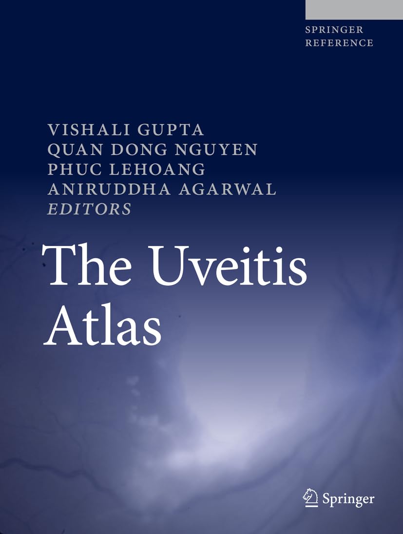
Introducing the comprehensive Uveitis Atlas, a must-have resource for ophthalmologists facing the diagnostic challenges of uveitic diseases. This atlas encompasses a wide range of intraocular conditions, providing valuable insights and diagnostic assistance for specialists in uveitis and general ophthalmologists.
Key features of the Uveitis Atlas include:
– Detailed descriptions and visual representations of uveitic entities
– Utilization of slit lamp and anterior segment photographs, fundus photographs, and ancillary investigations
– Incorporation of more than 1000 images to aid in diagnosis
– Case-based format for clear context and treatment planning
Ancillary investigations like fluorescein angiography, indocyanine green angiography, and optical coherence tomography, among others, play a pivotal role in reaching accurate diagnoses. Radiological imaging, systemic work-ups, and laboratory investigations are also crucial in establishing the root cause of uveitis.
With 136 chapters covering various uveitic conditions, this atlas serves as a go-to reference guide for practitioners seeking the most current information. Regular updates ensure that readers stay informed about new insights, techniques, and treatments relevant to uveitis management.
Whether you are a seasoned uveitis specialist or a general ophthalmologist, the Uveitis Atlas is designed to enhance your diagnostic skills and treatment approach. Stay ahead in the field of ophthalmology with this comprehensive resource.



Reviews
There are no reviews yet.