
By Julia Crim MD
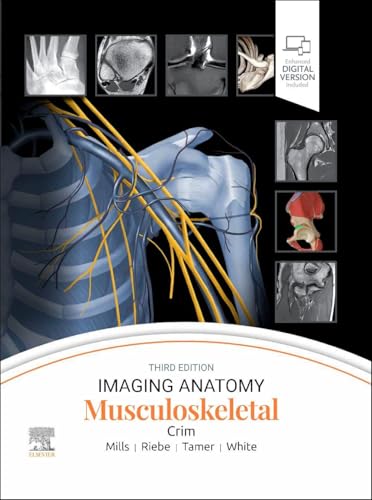
The third edition of Imaging Anatomy: Musculoskeletal is an essential reference for professionals in the medical imaging field. This comprehensive guide, authored by Dr. Julia Crim and her team of experts, offers new chapters and updated content, complemented by all-new 3T and 7T MR images that provide unparalleled detail of human anatomy. With over 1,900 high-quality images and illustrations, this anatomic atlas is meticulously designed for radiologists, orthopedic surgeons, sports medicine practitioners, and all specialists involved with musculoskeletal imaging.
- All-new 3T and 7T MR images deliver exceptional imaging detail, enhancing diagnostic accuracy.
- Features chapters on pediatric skeletal development, providing insights into growth and development.
- Includes radiographic, CT, and MR images supported by large, extensively labeled artist illustrations.
- New chapters cover the brachial plexus, scapula, clavicle, and sternum, expanding the scope of musculoskeletal practice.
- Introductory chapters illuminate bone maturation, bone marrow, and the musculotendinous unit.
- Overview chapters for each region offer essential information and expert medical illustrations, while focused chapters delve into complex anatomic issues such as the posterolateral corner of the knee.
- Detailed MR atlas chapters provide anatomy visualization in at least three planes.
- Discusses common normal variants, imaging pitfalls, and clinical applications for each area.
- Text updates reflect improved MR visualization of small structures and current clinical concerns related to anatomy.
- The book employs a time-saving, bulleted format for easy comprehension.









Reviews
There are no reviews yet.