
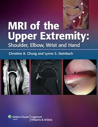
MRI of the Upper Extremity is an essential and comprehensive guide for medical professionals seeking to master MRI evaluation of the shoulder, elbow, wrist, hand, and finger disorders. This highly illustrated text/atlas provides a practical approach to MRI interpretation, focusing on the critical clinical correlations of imaging findings.
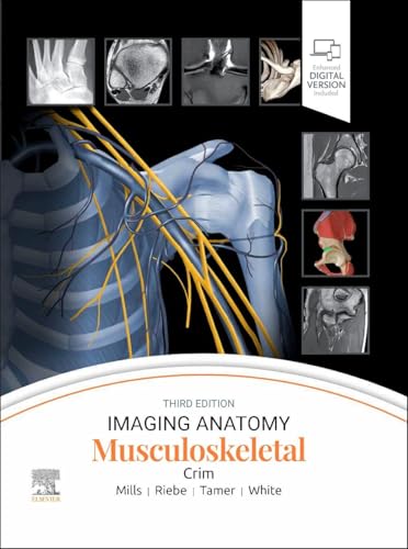
Featuring over 1,100 MRI scans, this book vividly displays both normal anatomy and pathologic findings. A full-color cadaveric atlas further enhances understanding by familiarizing readers with the anatomical structures seen on MR images.
Each joint’s section begins with a detailed review of MRI anatomy using cadaveric correlation, then transitions into technical MR imaging considerations and clinical assessments. Subsequent chapters delve into and illustrate MRI findings for specific disorders, including rotator cuff disease, nerve entrapment syndromes, osteochondral bodies, and triangular fibrocartilage disorders.
Who should read this book? This book is an invaluable resource for radiologists, orthopedic surgeons, and medical students who aim to deepen their understanding of MRI diagnostics in upper extremity disorders. Its practical approach and extensive illustrations make it a standout resource in the field of medical imaging.
What makes it special? The combination of extensive MRI scans along with a full-color cadaveric atlas offers a unique learning experience, making complex anatomical structures and disorders easier to understand and diagnose.
Discover the ultimate resource for MRI evaluation of the upper extremities and enhance your diagnostic skills with this authoritative guide.
Authors:
Christine B. Chung MD (Editor)
Edition:
1st
Publication Date:
November 5, 2009





Reviews
There are no reviews yet.