
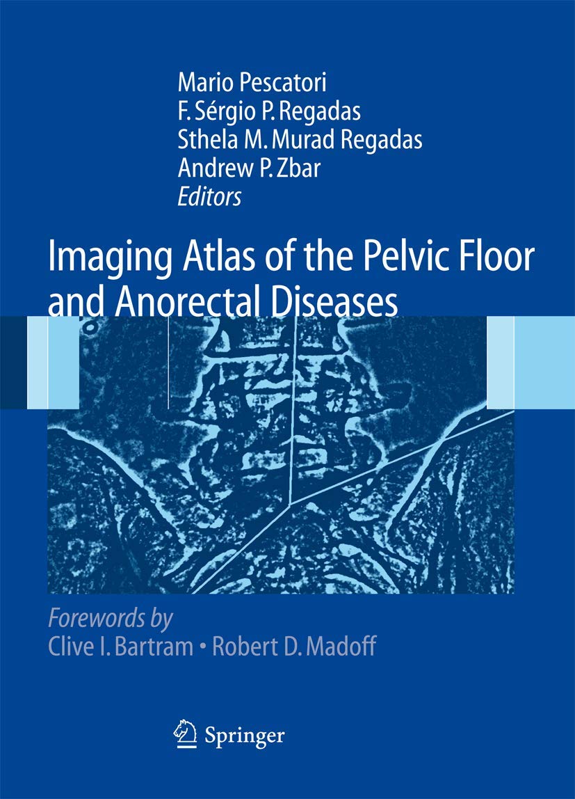
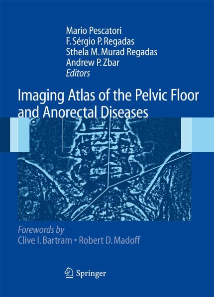
Discover the cutting-edge world of medical imaging in anorectal and pelvic floor disorders with this comprehensive atlas. As imaging technology advances, notably through three-dimensional ultrasound and magnetic resonance imaging (MRI), healthcare professionals can achieve high anatomical resolution and precise tissue differentiation. These innovations are crucial in the investigation and management of these complex conditions.
The atlas delves into the latest developments, such as three-dimensional endosonography for anorectal conditions and MRI for anal fistula. It also explores dynamic studies of the pelvic floor using both ultrasound and MRI, providing a holistic view of the field. Organized into four detailed sections—covering basic anatomy, anal/perianal disease, rectal/perirectal disease, and functional assessment—this atlas offers an easy-to-understand guide to modern imaging techniques.
One of the key challenges in creating an atlas is finding the right balance between text and images. This atlas excels at achieving this balance, offering exceptional images and diagrams alongside concise, informative text. The invited commentaries at the end of each chapter add valuable context, making the chapters more comprehensive and enriching the reader’s understanding.
Ideal for medical professionals, radiologists, and students, this atlas is an essential resource for those seeking to deepen their knowledge in the field of medical imaging. With its clear structure and detailed explanations, it is a must-have reference for anyone involved in the diagnosis and management of anorectal and pelvic floor disorders.
Enhance your understanding of this critical area in medical science by exploring this expertly curated atlas. Read more and elevate your expertise today!
Authors:
Mario
Edition:
2008th
Publication Date:
August 6, 2008

Reviews
There are no reviews yet.